Summary:
Achalasia cardia is a motility disorder of the lower esophageal sphincter, commonly presented with dysphagia. The reported case was presented with dysphagia & chest pain for 5 years. The patient was successfully managed by Laparoscopic cardiomyotomy with DOR fundoplication.
List of Authors:
- Dr. Mohammad Emrul Hasan Khan (Corresponding Author)
MBBS, FCPS (Surgery), MS (Hepatobiliary Surgery)
Associate Professor (Hepatobiliary & Pancreatic Surgery)
Dhaka Medical College, Dhaka
dremrul@gmail.com, Mobile No: 01713242529 - Dr. Zilanur Rahman
MBBS, MS (Hepatobiliary Surgery)
Surgery Specialist
Bangladesh Specialized Hospital, Shyamoli, Dhaka
Mobile No: 01714459049 - Dr. Masudur Rana Bhuiyan
MBBS, FCPS (Surgery)
Registrar (Surgical Gastroenterology)
Sheikh Russel National Gastroliver Institute & Hospital
Mobile No: 01717497101 - Dr. Shaila Sagor
MBBS, MS (General Surgery)
Assistant Professor
Bangladesh Institute of Health Science General Hospital, Dhaka
Mobile No: 01717495387
Introduction:
A primary benign motility disorder of esophagus, achalasia cardia develops due to non-relaxing lower esophageal sphincter (LES).1,2 It is commonly manifested by dysphagia for both solid (initial stage) and liquid (advance stage) diet, regurgitation, and chest pain.3,4 A rare condition occurring in 1 out of 10000 individuals, achalasia cardia is not well-managed with medicines.1 Non-surgical management like- use of calcium channel blockers, endoscopic guided pneumatic balloon dilatation or botulinum toxin injection have had significant treatment failure.4 Surgical treatment- Heller’s cardiomyotomy with anti-reflux procedure like DOR fundoplication improves the clinical outcome.4,5 Surgical treatment gained popularity only after the establishment of non-invasive laparoscopic Heller cardiomyotomy with DOR Fundoplication.1
Case Report:
A 16-year young boy presented with difficulty in deglutition for 5 years. During the initial period he was unable to eat solid food. Gradually, with time his problem worsened. At a later stage he became unable to take even soft foods and could only swallow small amounts of liquid, which is when he had to come for further intervention. Barium swallow X-Ray of esophagus & upper gastrointestinal (GI) endoscopy showed typical features of achalasia cardia. After proper preoperative evaluation and counseling, Laparoscopic Heller’s cardio myotomy with DOR Fundoplication was performed. On the day after the surgery the patient was allowed to take a soft diet and was discharged. On the 1st follow up at 7th post operative day (POD) he had no deglutition difficulties.
Operative Procedure:
Under general anesthesia the patient was placed in reverse Trendelenburg position. The surgeon stood in between the legs of the patient and the monitor was placed at the head end of table. 10mm optical port was made at supra-umbilical region in blind technique. All other ports were 5mm and made under direct visualization. 2 working ports were made 2cm above the optical port at the lateral border of rectus, 1 assistant port at left anterior axillary line at the same level and 1 epigastric port for liver retraction.
Dissection started from the right side of esophagus by cutting gastro-hepatic ligament using ligasure. The left hepatic branch from the left gastric artery was identified and secured. Dissection was proceeded through the avascular plane between the right crus and posterior wall of esophagus. Angle of His was detached and left crus of diaphragm was dissected from esophagus. About 6 to 8 cm of lower end of esophagus was mobilized from mediastinum. Anterior vagus nerve was identified and secured. Marking (5cm above and 1cm below the gastro-esophageal junction) was given using hook with monopolar diathermy. Myotomy was done bluntly by splitting the longitudinal fibers by using 2 meriland forceps. Circular fibers were cut by monopolar diathermy or by ligasure.

Once the myotomy was complete, about 50% of esophageal mucosa was bluntly separated from muscle edges. The nasogastric tube was pulled back into the distal esophagus and normal saline was injected and ensured that there was no mucosal perforation.
An anti-reflux procedure DOR fundoplication was done by overlapping the fundus of stomach anterior to esophagus. 3 interrupted sutures were placed on fundus, left crus, and left margin of esophageal muscle then another 3 stiches were given along fundus, right margin of esophageal muscle, and right crus. During the whole procedure, hemostasis was achieved by gauze pressure. Total operation time was about 110 minutes.
Post-operative care:
On 1st POD patient was allowed liquid followed by soft diet and discharged at 2nd POD with advice to take soft diet up-to 3 weeks. Normal diet was allowed after that but was prohibited from having fish or meat bone.
Discussion:
Achalasia cardia is a less common motility disorder of esophagus.4 As there are no ganglion cells in myenteric plexus, LES remain closed during swallowing.3,4 In early stage of the disease some food can pass beyond LES by powerful contraction of normal portion of esophagus.4 In later stage due to inflammation and neural fibrosis, contraction power is reduced, esophagus becomes dilated, and esophagus empties mainly by hydrostatic pressure of its content.4 So, most patients at early stage have dysphagia with solid food and in later stage they develop dysphagia even with liquid. Some patients also complaint of chest pain,4 which is due to inappropriate contraction of esophagus. At a later stage patient may be presented with regurgitation. Achalasia commonly occurs in middle age but can occur at any age.2
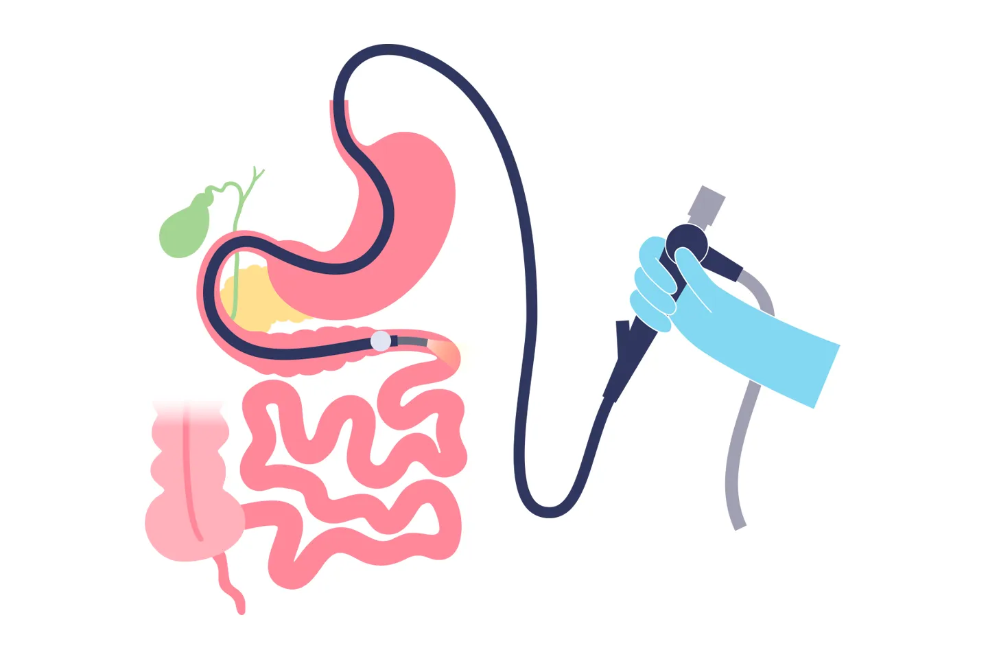
Barium swallow X-Ray of esophagus showing bird’s beak stricture at lower end with dilated proximal part is a diagnostic feature of achalasia cardia.4 Achalasia also may be suspected from upper GI endoscopy.4 A tight cardia and residual food at esophagus are the endoscopic features of achalasia.4 A firm diagnosis can be made by esophageal manometry.1,4
Among all the motility disorders of esophagus, achalasia responds well with treatment. Medical treatment by calcium channel blocker is ineffective for long term use.2,4 Botulinum toxin effect is not permanent and requires frequent re-treatment to maintain an efficacy rate of 65%.1,3 Pneumatic forceful dilatation is curative in 75-85% of cases.1,4 However, it is <50% effective in patients younger than 40 years of age and is rarely effective in adolescents.1,2,4 Heller’s myotomy with fundoplication is successful in 90-95% of cases.2,4 Despite these excellent results, historically 75% of patients opted for balloon dilation as primary therapy when both options were presented elaborately.1 Laparoscopic surgery being a non-invasive procedure causing less pain, almost no scarring, and the shortest downtime in postoperative disability has made surgical therapy more attractive to patients.1,4
In this case report, the patient was presented with typical features of achalasia cardia. Barium swallow X-Ray and upper GI endoscopy found characteristic findings of achalasia. As the patient’s age was 16 years, laparoscopic Heller’s cardiomyotomy with DOR fundoplication was the preferred treatment option. Right after one day of surgery, the patient could take a soft diet and now can eat normal food.
Conclusion:
Laparoscopic cardiomyotomy with DOR fundoplication is a safe, effective procedure for the treatment of achalasia. Outcome is very promising, especially in adolescents.
References:
- John G. Hunter., Thadeus L. Trus., Gene D. Branum., J. Patrick Waring. Laparoscopic Heller Myotomy and Fundoplication for Achalasia. Annals of surgery 1997; 225 (6), 655-665.
- G Ramacciato., FA D’Angelo., P Aurello., M Del Gaudio., G Varotti., P Mercantini., R Bellagamba., G Ercolani. Laparoscopic Heller myotomy with or without partial fundoplication: A matter of debate. World Journal of Gastroenterology 2005; 11(10):1558-1561.
- Damian Mayo., Ewen A. Griffiths., Omar A. Khan., Mark A. Szymankiewicz., Christian W. Wakefield., Sarah K. Thompson. Does the Addition of a Fundoplication Improve Outcomes for Patients Undergoing Laparoscopic Heller’s Cardiomyotomy? International Journal of Surgery 2012; 10 (2012): 301-304.
- Derek Elderson. The esophagus. Bailey & Love’s short practice of Surgery 2018; 27(62), 1067-1105.
- Ackroyd R, Watson DI, Devitt PG, Jamieson GG. Laparoscopic cardiomyotomy and anterior partial fundoplication for achalasia. Surg Endosc 2001; 15: 683-686