
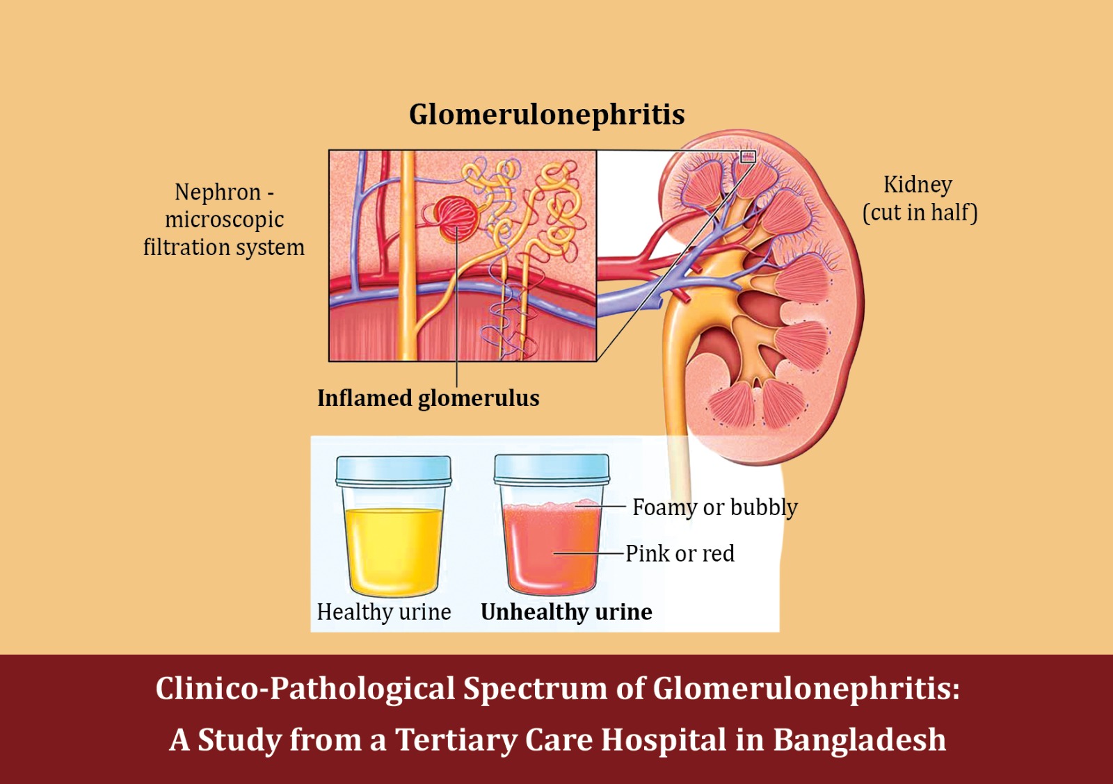

Abstract:
Background: Glomerulonephritis (GN) is a significant cause of chronic kidney disease, yet evidence from Bangladesh remains limited. This study aimed to describe the clinicopathological spectrum of GN among patients undergoing native kidney biopsy at a tertiary hospital.
Methods: A retrospective review was conducted at Sher-E-Bangla Medical College, Barisal, Bangladesh, including 67 patients aged ≥12 years with biopsy-proven GN. Demographic, clinical, laboratory, and histopathological data were analyzed using descriptive statistics.
Results: The median age of the patients was 30 years (IQR: 21–47), with 40 patients (59.7%) being female. Most patients (86.6%) had a normal body mass index (BMI). Diabetes mellitus and hypertension were present in 4.5% and 13.4% of the patients, respectively. Nephrotic presentations occurred in 38 patients (56.7%), while nephritic presentations were observed in 29 patients (43.3%). The median serum creatinine level was 1.02 mg/dL, and the estimated glomerular filtration rate (eGFR) was 80 mL/min/1.73 m². Hypoalbuminemia (2.5 g/dL), hypercholesterolemia (321 mg/dL), and proteinuria (4.8 g/day) were common findings. Low complement C3 and C4 levels were noted in 48.5% and 46.9% of the patients, respectively. Antinuclear antibody (ANA) was positive in 28.9% of cases, anti-double-stranded DNA (anti-dsDNA) in 36.4%, and anti-neutrophil cytoplasmic antibody (ANCA) in 40% (2 out of 5). Histopathology revealed focal segmental glomerulosclerosis (FSGS) (22.4%) and lupus nephritis (22.4%) as the most common diagnosis, followed by minimal change disease (MCD) (13.4%), membranoproliferative glomerulonephritis (MPGN) (10.4%), and infection-related glomerulonephritis (11.9%). Nephrotic presentations predominated in FSGS (36.8%) and MCD (15.8%), while nephritic presentations were more frequent in lupus nephritis (34.4%) and infection-related glomerulonephritis (21.9%).
Conclusion: In this cohort, focal segmental glomerulosclerosis and lupus nephritis were the leading causes of GN. The distinct clinicopathological correlations emphasize the diagnostic value of renal biopsy and highlight the need for larger studies in Bangladesh.
Keywords: Glomerulonephritis, Chronic Kidney Disease, Bangladesh, Clinicopathological Spectrum.
Introduction
Glomerulonephritis (GN) remains a major contributor to the global burden of chronic kidney disease (CKD), a condition that affected roughly 650–700 million people worldwide and accounted for more than 1.2 million deaths in 2017; unlike other noncommunicable diseases, CKD mortality has risen over recent decades and is projected to climb further in global rankings of causes of death.1, 2 Although the specific proportion of CKD directly attributable to GN varies by region and biopsy practice, population and registry data consistently show that immune-mediated glomerular diseases (e.g., IgA nephropathy, membranous nephropathy, lupus nephritis, infection-related GN, and complement-mediated disease) are key drivers of proteinuria, hematuria, progressive loss of kidney function, and dialysis initiation across age groups.3, 4 The epidemiology of GN is dynamic: for example, acute post-streptococcal GN has declined in many high-income settings while staphylococcus-associated and other infection-related forms have become more prominent in adults, often with worse renal outcomes.5–7 Recent Global Burden of Disease analyses of acute GN suggest declines in incidence and disability-adjusted life years since 1990, yet the absolute numbers and clinical consequences remain substantial.8 High-quality clinic-pathological correlation is central to the diagnosis and management of GN. Histopathology refines or revises clinical impressions, assigns disease class (e.g., ISN/RPS classes in lupus nephritis), grades activity and chronicity, and guides immunosuppression and supportive care; in turn, clinical context (syndrome at presentation, serologies, complements, comorbid infection) is essential to interpret biopsy findings and to anticipate prognosis.9, 10 Contemporary international guidelines (KDIGO 2021, with ongoing updates) emphasize integrating kidney biopsy patterns with clinical and laboratory features to individualize therapy and to avoid both under- and overtreatment.10, 11 This integration is particularly important where infection-related or complement-mediated processes may mimic primary GN and where timely recognition changes antimicrobial, immunosuppressive, and supportive strategies.12 Despite the clinical importance of GN, Bangladeshi data remain limited and fragmented. There is no national kidney biopsy registry, and available reports are largely single-center series with heterogeneous inclusion criteria and time frames, constraining inferences about national patterns and trends.13 Existing local studies many focused on specific entities such as lupus nephritis underscore the value of clinicopathological correlation but are small and not population-based, further highlighting evidence gaps.14, 15 Moreover, Bangladesh shares regional risk modifiers (high infectious disease burden, variable access to nephrology and pathology services, and late presentation) that can shift GN spectra toward infection-related disease and advanced chronicity at biopsy, yet systematic documentation is sparse.5, 16 As health systems plan for escalating CKD care needs, up-to-date granular descriptions of GN phenotypes, serologic profiles (e.g., complements, autoantibodies), and initial clinical syndromes (nephrotic vs nephritic) in Bangladeshi cohorts are urgently required. This study aims to describe the clinic-pathological spectrum of glomerulonephritis (GN) among patients undergoing native-kidney biopsy at a tertiary care hospital in Bangladesh. Specifically, it seeks to (i) standardize histopathological diagnosis into contemporary categories, (ii) characterize presenting clinical syndromes and laboratory profiles, and (iii) explore basic clinicopathological correlations across diagnostic groups. By generating rigorously categorized data from a real-world Bangladeshi biopsy cohort, the study addresses a critical evidence gap and provides foundational data for future multi-center registries, in alignment with KDIGO’s call for context-specific application of guideline principles in glomerular diseases.
Methods
Study Design and Sefling
This study employed a retrospective chart review design, conducted at Sher-E-Bangla Medical College, Barisal, Bangladesh, a tertiary care referral center in Bangladesh. The hospital serves a diverse patient population from both urban and rural catchment areas, providing comprehensive nephrology services including kidney biopsy, serological evaluation, and longitudinal follow-up.
Study Population and Sample Size
The study included 67 patients with biopsy-proven glomerulonephritis (GN), diagnosed between June 2024 to June 2025 at Barishal Medical College Hospital. Eligible patients were aged 12 years and above and had undergone native kidney biopsy with complete clinical and laboratory documentation. Patients were excluded if their records were incomplete, if they had secondary renal disease unrelated to GN, or if the biopsy sample was inadequate for definitive histopathological diagnosis.
Data Collection and Variables
Demographic, clinical, and laboratory data were systematically extracted from patient charts and biopsy reports. Demographic variables included age, sex, weight, and height. Renal function parameters comprise serum creatinine and estimate glomerular filtration rate (eGFR). The urinary parameters collected included 24-hour proteinuria, and urinalysis, which encompassed red blood cell count and hematuria. Biochemical and serological data included serum albumin, cholesterol, complement levels (C3, C4), antinuclear antibody (ANA), anti-double-stranded DNA (anti-dsDNA), and anti-neutrophil cytoplasmic antibody (ANCA), which consists of two main types: C-ANCA and P-ANCA. Information on comorbidities and clinical features such as diabetes mellitus, hypertension, edema, high-color urine, oliguria, nephrotic versus nephritic presentation, and prior history of infection was also recorded. Histopathological findings were standardized into recognized categories, including minimal change disease (MCD), focal segmental glomerulosclerosis (FSGS) variants, membranous nephropathy, lupus nephritis classes II–V, IgA and IgM nephropathies, membranoproliferative GN (MPGN), complement-mediated GN (C3 glomerulopathy), infection-related GN, diabetic nephropathy, and ANCA-associated vasculitis. All data were anonymized and entered a structured electronic database to ensure accuracy, consistency, and ease of analysis.
Results
Demographic and Clinical Characteristics
The median age of participants was 30 years, with an interquartile range (IQR) of 21 to 47 years. Among them, 27 (40.3%) were male and 40 (59.7%) were female. The mean weight was 55.0 ± 6.9 kg, and the mean height was 155.0 ± 8.7 cm. Regarding BMI, 2 patients (3.0%) were underweighted, 58 (86.6%) had
normal BMI, 6 (9.0%) were overweight, and 1 (1.5%) was obese. Diabetes mellitus was present in 3 patients (4.5%), while hypertension was recorded in 9 patients (13.4%). In terms of edema, 5 patients (7.5%) had no edema, 17 (25.4%) had mild edema, 19 (28.4%) had
moderate edema, 25 (37.3%) had severe edema, and 1 (1.5%) had very severe edema. A nephrotic presentation was observed in 38 patients (56.7%), while a nephritic presentation was found in 32 patients (47.8%). A history of infection was reported in 4 patients (6.0%). Oliguria was present in 11 patients (16.4%), and high-colour urine was documented in 8 patients (11.9%) (Table 1).
Table 1: Demographic and Clinical Characteristics of the Patients with Biopsy-Proven Glomerulonephritis (N = 67)
| Variable | Value |
| Age, years, median (IQR) | 30 (21 – 47) |
| Sex, n (%) | |
| Male | 27 (40.3) |
| Female | 40 (59.7) |
| Weight, kg, mean ± SD | 55.0 ± 6.9 |
| Height, cm, mean ± SD | 155.0 ± 8.7 |
| BMI Classification, n (%) | |
| Underweight (<18.5) | 2 (3.0) |
| Normal (18.5–24.9) | 58 (86.6) |
| Overweight (25–29.9) | 6 (9.0) |
| Obese (≥30) | 1 (1.5) |
| Diabetes Mellitus, n (%) | 3 (4.5) |
| Hypertension, n (%) | 9 (13.4) |
| Edema, n (%) | |
| Nil | 5 (7.5) |
| Mild (1+) | 17 (25.4) |
| Moderate (2+) | 19 (28.4) |
| Severe (3+) | 25 (37.3) |
| Very severe (4+) | 1 (1.5) |
| Nephrotic presentation, n (%) | 38 (56.7) |
| Nephritic presentation, n (%) | 32 (47.8) |
| History of infection, n (%) | 4 (6.0) |
| Oliguria, n (%) | 11 (16.4) |
| High-color urine, n (%) | 8 (11.9) |
Laboratory Characteristics of Patients
Among the 67 patients with biopsy-proven glomerulonephritis, the median serum creatinine was 1.02 mg/dL (IQR: 0.9–1.3), with a corresponding median eGFR of 80 mL/min/1.73 m² (IQR: 55–116). Serum albumin had a median of 2.5 g/dL (IQR: 2–3.1). The median serum cholesterol level was 321 mg/dL (IQR: 210–500). Urinary parameters showed a median proteinuria of 4.8 g/day (IQR: 3.1–8.2) and a median of 3 urine RBCs/hpf (IQR: 0–11). Complement levels demonstrated that 16 patients (48.5%) had low serum C3, while 17 patients (51.5%) had normal C3. Similarly, 15 patients (46.9%) had low serum C4, compared with 17 patients (53.1%) who had normal levels. Regarding serological markers, 13 of 45 patients (28.9%) tested positive for ANA, 12 of 33 patients (36.4%) were positive for anti-DNA, and 2 of 5 patients (40.0%) were positive for ANCA (Table 2).
Table 2: Laboratory Characteristics of Patients with Biopsy-Proven Glomerulonephritis (n = 67)
| Variable | Median (IQR) / n (%) |
| Serum creatinine, mg/dL | 1.02 (0.9–1.3) |
| eGFR, mL/min/1.73 m² | 80 (55–116) |
| Serum albumin, g/dL | 2.5 (2–3.1) |
| Serum cholesterol, mg/dL | 321 (210–500) |
| Urine protein (g/day) | 4.8 (3.1–8.2) |
| Urine RBCs/hpf | 3 (0–11) |
| Serum C3, mg/dL, n (%) | |
| Low | 16 (48.5) |
| Normal | 17 (51.5) |
| Serum C4, mg/dL, n (%) | |
| Low | 15 (46.9) |
| Normal | 17 (53.1) |
| ANA positive, (n = 45, %) | 13 (28.9) |
| Anti-DSDNA positive, (n = 33, %) | 12 (36.4) |
| ANCA positive, (n = 5, %) | 2 (40.0) |
IQR: inter-quartile range, eGFR: estimated glomerular filtration rate, RBC: red blood cell, hpf: high-power field, GN indicates membranoproliferative, IgA indicates Immunoglobulin A, ANA: Antinuclear antibody, Anti-dsDNA: anti-double-stranded DNA, ANCA: anti-neutrophil cytoplasmic antibody.
Histopathological Spectrum of Glomerulonephritis Among the 67 patients with biopsy-proven glomerulonephritis, Minimal Change Disease (MCD) was observed in 9 cases (13.4%). Focal Segmental Glomerulosclerosis (FSGS, all variants) was identified in 15 cases (22.4%). Membranous Nephropathy was reported in 5 cases (7.5%), while Membranoproliferative Glomerulonephritis (GN) was detected in 7 cases (10.5%). Lupus Nephritis (Classes II–V) was found in 15 cases (22.4%). Infection-Related GN was noted in 8 cases (11.9%), and Immunoglobulin A (IgA) Nephropathy was observed in 3 cases (4.5%). Diabetic Nephropathy and ANCA-Associated Vasculitis were each identified in 2 cases (3.0%) (Table 3).
Table 3: Histopathological Spectrum of Glomerulonephritis (n = 67)
| GN Category | n (%) |
| MCD | 9 (13.4) |
| FSGS (all variants) | 15 (22.4) |
| Membranous Nephropathy | 5 (7.5) |
| Membranoproliferative GN | 7 (10.5) |
| Lupus Nephritis (II–V) | 15 (22.4) |
| Infection-Related GN | 8 (11.9) |
| IgA Nephropathy | 3 (4.5) |
| Diabetic Nephropathy | 2 (3.0) |
| ANCA-Associated Vasculitis | 2 (3.0) |
GN: membranoproliferative, MCD: minimal change disease, IgA: immunoglobulin A; FSGS: focal segmental glomerulosclerosis. ANCA: anti-neutrophil cytoplasmic antibody; FSGS (all variants) includes not otherwise specified, tip lesion, perihilar, and other variants
Clinicopathological Correlation: Presentation and GN Category
Among patients with nephrotic presentation, the most frequent histopathological diagnosis was FSGS (14 cases, 36.8%), followed by Minimal Change Disease (MCD) (6 cases, 15.8%). Lupus nephritis (II–V) and membranoproliferative GN were each observed in 5 (13.2%) and 4 (10.5%) cases, respectively. Membranous nephropathy accounted for 4 cases (10.5%), while diabetic nephropathy was noted in 2 cases (5.3%). Infection-related GN and IgA nephropathy were found in 2 (5.3%) and 1 (2.6%) case, respectively. No case of ANCA-associated vasculitis presented with nephrotic syndrome. In patients with nephritic presentation, the leading diagnosis was lupus nephritis (II–V) (11 cases, 34.4%), followed by infection-related GN (7 cases, 21.9%). Membranoproliferative glomerulonephritis (GN) was observed in 3 cases (9.4%). In contrast, minimal change disease (MCD), focal segmental glomerulosclerosis (FSGS), IgA nephropathy, and ANCA-associated vasculitis each accounted for 2 cases (6.3%) in their respective categories. Membranous nephropathy and diabetic nephropathy were less frequent, with 1 case (3.1%) each (Table 4).
Table 4: Clinicopathological Correlation: Presentation and GN Category
| GN Category | Nephrotic (n = 38, %) | Nephritic (n = 32, %) |
| MCD | 6 (15.8) | 2 (6.3) |
| FSGS (all variants) | 14 (36.8) | 2 (6.3) |
| Membranous Nephropathy | 4 (10.5) | 1 (3.1) |
| Membranoproliferative GN | 4 (10.5) | 3 (9.4) |
| Lupus Nephritis (II–V) | 5 (13.2) | 11 (34.4) |
| Infection-Related GN | 2 (5.3) | 7 (21.9) |
| IgA Nephropathy | 1 (2.6) | 2 (6.3) |
| Diabetic Nephropathy | 2 (5.3) | 1 (3.1) |
| ANCA-Associated Vasculitis | 0 (0.0) | 2 (6.3) |
MCD: minimal change disease, GN: membranoproliferative; FSGS: focal segmental glomerulosclerosis, IgA: immunoglobulin A. ANCA: anti-neutrophil cytoplasmic antibody; FSGS (all variants) includes not otherwise specified, tip lesion, perihilar, and other variants
Discussion
The study population had a median age of 30 years, indicating a predominance of younger adults. Females constituted the majority (59.7%) compared to males (40.3%), which is consistent with prior observations that certain glomerular diseases, such as lupus nephritis, are more frequent among females.17 The mean body weight and height were 55.0 ± 6.9 kg and 155.0 ± 8.7 cm, respectively, with most participants maintaining a normal BMI (86.6%). Comorbid diabetes mellitus and hypertension were relatively uncommon, present in 4.5% and 13.4% of patients, respectively, highlighting that the cohort largely comprised patients without significant metabolic risk factors. Edema was observed in nearly all patients, ranging from mild (25.4%) to severe (37.3%), reflecting the frequent nephrotic involvement in the cohort. Nephrotic syndrome was the predominant clinical presentation (56.7%), while nephritic syndrome occurred in 47.8% of patients, indicating an overlap in clinical manifestations of biopsy-proven glomerulonephritis.9 History of infection, oliguria, and high-color urine were less common, reported in 6.0%, 16.4%, and 11.9% of cases, respectively.
Laboratory evaluation revealed a median serum creatinine of 1.02 mg/dL and a median eGFR of 80 mL/min/1.73 m², suggesting that most patients had preserved renal function at presentation. Serum albumin was notably low (median 2.5 g/dL), while serum cholesterol was elevated (median 321 mg/dL), consistent with nephrotic-range proteinuria (median 4.8 g/day).18 Hematuria was variable, with a median of 3 RBCs/hpf. Complement levels were reduced in nearly half of the patients for both C3 (48.5%) and C4 (46.9%), reflecting possible immune-complex–mediated pathology. ANA and anti-DSDNA positivity rates (28.9% and 36.4%, respectively) aligned with the observed prevalence of lupus nephritis in this cohort, whereas ANCA positivity was limited to 2 of 5 tested patients (40%), consistent with the low frequency of ANCA-associated vasculitis.19 Histopathologic ally, FSGS (22.4%) and lupus nephritis (22.4%) were the most common diagnosis, followed by minimal change disease (13.4%) and membranoproliferative GN (10.4%). Membranous nephropathy, infection-related GN, IgA nephropathy, diabetic nephropathy, and ANCA-associated vasculitis were less frequent, consistent with patterns reported in other regional and international series.20, 21 Clinicopathological correlation demonstrated that nephrotic syndrome was most frequently associated with FSGS (36.8%) and MCD (15.8%), whereas nephritic presentations were predominantly linked to lupus nephritis (34.4%) and infection-related GN (21.9%). Membranoproliferative GN, IgA nephropathy, and ANCA-associated vasculitis contributed modestly to both presentations. Membranous nephropathy and diabetic nephropathy were rarely associated with nephritic features. These findings corroborate prior observations that FSGS and MCD commonly present with proteinuria-dominant nephrotic syndrome, while lupus nephritis and infection-related GN are more likely to manifest with hematuria and nephritic features.22,23 Overall, the demographic, clinical, laboratory, and histopathological profile of this cohort underscores the heterogeneity of glomerular diseases in adults, highlighting the importance of renal biopsy for accurate diagnosis and guiding management strategies.
Conclusion
In this cohort of adults with biopsy-proven glomerulonephritis, the majority were young adults with preserved renal functions at presentation. Focal Segmental Glomerulosclerosis and lupus nephritis were the most prevalent histopathological diagnosis. Nephrotic syndrome was predominantly associated with FSGS and Minimal Change Disease, whereas nephritic presentations were primarily linked to lupus nephritis and infection-related glomerulonephritis. Complement abnormalities and serological markers were consistent with underlying immune-mediated pathology in a substantial proportion of patients. These findings highlight the heterogeneity of clinical and histopathological presentations, emphasizing the critical role of renal biopsy in guiding accurate diagnosis and management strategies for glomerular diseases.
Author of this Article
Dr. Md Hafizur Rahman
Assistant Professor,
Department of Nephrology, Sher-E-Bangla Medical College, Barisal, Bangladesh
References
1. Xie K, Cao H, Ling S, Zhong J, Chen H, Chen P, et al. Global, regional, and national burden of chronic kidney disease, 1990-2021: a systematic analysis for the global burden of disease study 2021. Front Endocrinol. 2025 Mar 5; 16:1526482.
2. Deng L, Guo S, Liu Y, Zhou Y, Liu Y, Zheng X, et al. Global, regional, and national burden of chronic kidney disease and its underlying etiologies from 1990 to 2021: a systematic analysis for the Global Burden of Disease Study 2021. BMC Public Health. 2025 Feb 17;25(1):636.
3. Thomé GG, Bianchini T, Bringhenti RN, Schaefer PG, Barros EJG, Veronese FV. The spectrum of biopsy-proven glomerular diseases in a tertiary Hospital in Southern Brazil. BMC Nephrol. 2021 Dec 13; 22:414.
4. Schena FP, Nistor I. Epidemiology of IgA Nephropathy: A Global Perspective. Semin Nephrol. 2018 Sep;38(5):435–42.
5. Satoskar AA, Parikh SV, Nadasdy T. Epidemiology, pathogenesis, treatment and outcomes of infection-associated glomerulonephritis. Nat Rev Nephrol. 2020 Jan;16(1):32–50.
6. Medjeral-Thomas NR, Pusey CD. New Insights into Epidemiology and Outcome of Bacterial Infection–Related Glomerulonephritis. Clin J Am Soc Nephrol CJASN. 2021 Aug;16(8):1149–51.
7. Nasr SH, Radhakrishnan J, D’Agati VD. Bacterial infection–related glomerulonephritis in adults. Kidney Int. 2013 May 1;83(5):792–803.
8. Jiao Y, Chen X, Zhang T, Ma C, Shen C, Yu B. Global, regional, and national burden of acute glomerulonephritis from 1990 to 2021 and future trend predictions until 2036: a systematic analysis using the Global Burden of Disease Study 2021. Front Public Health ;13.
9. Rovin BH, Adler SG, Barratt J, Bridoux F, Burdge KA, Chan TM, et al. KDIGO 2021 Clinical Practice Guideline for the Management of Glomerular Diseases. Kidney Int. 2021 Oct 1;100(4): S1–276.
10. Rovin BH, Adler SG, Barratt J, Bridoux F, Burdge KA, Chan TM, et al. Executive summary of the KDIGO 2021 Guideline for the Management of Glomerular Diseases. Kidney Int. 2021 Oct;100(4):753–79.
11. Glomerular Diseases (GD) – KDIGO.
12. Khalighi MA, Chang A. Infection-Related Glomerulonephritis. Glomerular Dis. 2021 Apr 28;1(2):82–91.
13. Islam MdN. The Spectrum and Trends of Glomerulonephritis in Bangladesh: A Single Center Study. Public Health Open Access
14. Mubarak M. Lupus nephritis in Bangladesh: unfolding the story. J Ethnopharmacology. 2016 Jan 2;5(1):26–7.
15. Baqui MN, Akhter S, Kabir E, Islam MS. A clinicopathological study on lupus nephritis; experience of 34 cases from Bangladesh. J Ethnopharmacology. 2015 Jul 22;5(1):19–23.
16. Mohapatra A, Kakde S, Annapandian VM, Valson AT, Duhli N, Korula A, et al. Spectrum of biopsy proven renal disease in South Asian children: Two decades at a tropical tertiary care center. Nephrol Carlton Vic. 2018 Nov 1;23(11):1013–22.
17. Anders HJ, Saxena R, Zhao MH, Parodis I, Salmon JE, Mohan C. Lupus nephritis. Nat Rev Dis Primer. 2020 Jan 23;6(1):7.
18. Tapia C, Bashir K. Nephrotic Syndrome.
19. Bolotin D, O’Brien C, Ranganath VK, Kermani TA. ANCA-associated vasculitis in patients with rheumatoid arthritis: A single-center cohort study. Semin Arthritis Rheum. 2025 Apr 1; 71:152648.
20. Nasr SH, Sirac C, Bridoux F, Javaugue V, Bender S, Rinsant A, et al. Heavy Chain Fibrillary Glomerulonephritis: A Case Report. Am J Kidney Dis Off J Natl Kidney Found. 2019 Aug;74(2):276–80.
21. Prasad N, Yadav AK, Kundu M, Sethi J, Jaryal A, Sircar D, et al. Prescription Practices in Patients with Mild to Moderate CKD in India. Kidney Int Rep. 2021 Sep;6(9):2455–62.
22. Rout P, Hashmi MF, Baradhi KM. Focal Segmental Glomerulosclerosis. In: StatPearls. Treasure Island (FL): StatPearls Publishing; 2025
23. Rodríguez-Iturbe B, Burdmann EA, Barsoum RS. Glomerular Diseases Associated with Infection. Compr Clin Nephrol. 2010;662–7.