Abstract:
Fetus in fetu is a rare developmental abnormality in which a mass of tissue resumbling a fetus forms inside the body of its twin. It is a parasitic twin of a diamniotic, monozygotic twin. It should be differentiated from teratoma which has no axial arrangement. This operation is a challenging one as it adherent with major abdominal organ like small gut, pancreas, kidney etc. which may be injured during operation. Here we report a case of fetus in fetu (double fetus) which is the 1st case seen in Bangladesh.
Introduction:
Fetas in feta is one of the rarest cases among intraadominal mass in children. It is difficult to differentiated from retroperitoneal teratoma. We present the diagnosis and management of a case fetus in fetu (double) in a 22 months old female child. A fetus in fetu is a monochorionic diamniotic monizygotic twin of its bearer. It is usually intraperitoneal or retroperitoneal but may also present in scrotum or in cranial cavity.1 It is usually surrounded by a membrane to analogous to amniotic sac and supplied by a single feeding vessel. A true placenta is usually absent. Absence of an independent circulatory system explains the subsequent growth retardation. Developmentally speaking it has gone through the stage of primitive streak which is why it has vertebral body and organ arranged around axis. This differentiates it from fetiform teratoma. Fetus in fetu is usually mal-formed because of pressure exerted by the host organ.2 This pathology is rare and the incidence is 1 per 500,000 birth. 3 Fetus in fetu is a rare condition with less than 200 cases reported in the world to the best of our knowledge.4 The present study is an effort to present a rare case of fetus in fetu with review of the available literature.
Case report:
A 22 months old girl was send to my chamber by my pediatrician teacher from his private chamber. Initially she was taken by her parents to Kaukhali health Complex of Rangamati District with the complaints of abdominal pain and hard mass in abdomen where they gave her symptomatic treatment and referred her to Chattagram Medical college Hospital. Instead of going to CMCH, parents brought her to Prof. Dr. Mesbahul Haque’s chamber. He examined her and told them to meet with a paediatric surgeon by mentioning My name.
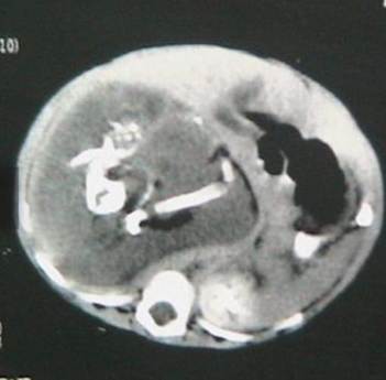
Her parents complained about a gradually increasing hard lump in her upper abdomen since last 6 months which was extending upto pelvis with occasional pain. There was no maternal illness, exposure to radiation or drug intake during pregnency. There was no family history of multiple pregnencies. There was no other gastrointestinal or genitourinary symptom. At Clinical examination a, bulky, firm, rounded mass of 14cm×16cm was palpated in Rt hypochondriac, epigastrium & umbilical region. The plain abdominal radiograph revealed a right upper quadrant mass containing calcified densities identifiable as fetal parts. CT Scan of abdoman with oral and intravenous contrast reveals a large retroperitoneal soft tissue mass with cystic component which is separated from Rt. Kidney. Her haemoglobin level was optimum and other haematological parameters were within normal limit.
Laparotomy was performed through a supraumbilical transverse incision. At surgery, a thick walled solido cystic mass was found in the retroperitoneal region which compressed IVC and duodenum and small gut were displaced towards left side. The mass enveloped entirely by a saclike capsule withan umbilical cord. The cord connected the mass to the branch of abdominal aorta.
The mass removed entirely through sharp & blant dissection after ligating of blood vs. and sections of cord. The mass was adherent with pancreas lightly which was gently separated. Abdomen was closed in layers with a drain tube in situ.
The patient recovered nicely and no ICU support was needed. Post operative period was uneventful and drain tube removed after 48 hours and feeding given after 24 hrs. Patient was discharged home on 4th POD.
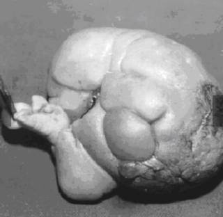
By opening the capsule, a fetiform structure was observed. Symmatrical, well developed four lower limbs with structure resembling toes were present. Rudimentory upper limbs present but it was 3 in members. Two heads present with hair.
Discussion :
Fetus in fetu is a usually single, however possibly multiple (here 2 in number) aberration of monozygotic, diamniotic twining in which unequal division of the totipotent inner cell mass of the developing blastocyst leads to the inclusion of a smaller cell mass within a maturing sister embryo.5 Thakral et al.6 reported equal male and female prevalence but Patankar et al. 7 and Federici et al. 8 noted a 2:1 male predominance. The common presentation of fetus in fetu is mass most commonly in the abdomen, almost 80% in the retroperitoneum. 9 The fetus in fetu produces symptoms due to mass effect leading to distension, difficulty in feeding, vomiting, pain and urinary retention.
In this case, during surgery, the fetus was found to be surrounded by a capsule that grossly ressmbled a fetal membrane. It contained a variable amount of fluid and was connected by a peduncle to a vascular structure of the host twin. Preoperative diagnosis is possible by radiograph, ultrasonography and computer tomograph. Though a rare anomaly, fetus in fetu can be identified radiologically in the pre-operative period.10 Radiological differential diagnosis included teratoma and meconium pseudocyst. 11 Pathological controversy arises during differentiation of fetus in fetu from a mature or well organized teratoma. According to willis,12 the presence of axial skeleton with vertebral axis and an appropriate arrangement of other limbs and organ goes more in favour of fetus in feta. Consistent with the theory of Willis, in my case, the vertebral column was detected by the radiologist. It was radio opaque as it was relatively calcified. However review of literature showed that in about 9% of case of fetus in fetu, no vertebral column was found even on radiological examination. 13
Therefore, Gonzalez- crassi suggested fetus in fetu to the applied to any structure in which the fetal form has a highly developed organogenesis or to the presence of vertebral axis. Our case did not fall in the first category at we observed the vertebral column in our fetus in fetu. On the other hand, teratoma is an accumalation of Pluripotent cell in which there is neither organogenesis nor vertebral segmentation,14 Another important aspect of fetus in fetu is that they neger become malignant whereas teratoma is known to become malignant.
Treatment of fetus in fetu is essentially surgical and excision give complete recovery. In our case, we completely resected the muss with no subsequent complication to the best of our knowledge. It is necessary to keep the child in follow up and surveillance as malignant recurrance has also been described.
CONCLUSIONS
Fetus in fetu is a rare and interesting entity that typically presents as an abdominal mass in infancy or early childhood. It can be diagnosed in the preoperative period.
Complete excision of mass is curative and confirmatory. Though a rare entity, it should be kept in mind as a differential diagnosis for lump abdomen in infancy and early childhood and should be well differentiate from teratoma which a common variant.
ACKNOWLEDGMENTS
We certify that this case report has not been previously published, nor is it under consideration by another journal. The authors have read this report thoroughly and to ensure honest work.
Author of this article
Prof. Dr. Nandan Kumar Mojumder
MCPS, MS (Pediatric Surgery)
Pediatric & Laparoscopic Surgeon
Professor, Southern Medical Collage and Hospital, Chottagram.
REFERENCES
1. Sutthiwan P, Sutthiwan I, Tree-trakan T. Fetus in fetu. J Pediatr Surg. 1983;18(3):290-2. doi: 10.1016/s0022-3468(83)80105-5. [DOI] [PubMed] [Google Scholar]
2. Du Plessis JP, Winship WS, Kirstein JD. Fetus in fetu and teratoma. A case report and review. S Afr Med J. 1974;48(50):2119-22. [PubMed] [ Google Scholar]
3. Hopkins KL, Dickson PK, Ball TI, Ricketts RR, O’Shea PA, Abramowsky CR. Fetus-in-fetu with malignant recurrence. J Pediatr Surg. 1997;32(10):1476-9. doi: 10.1016/s0022-3468(97)90567-4. [DOI] [PubMed] [Google Scholar]
4. Arlikar JD, Mane SB, Dhende NP, Sanghavi Y, Valand AG, Butale PR. Fetus in fetu: two case reports and review of literature. Pediatr Surg Int. 2009;25(3):289-92. doi: 10.1007/s00383-009-2328-8. [DOI] [PubMed] [Google Scholar] 5. Lewis RH. Fetus in fetu and retroperitoneal teratoma. Arch Dis Child. 1960;36:220-6. doi: 10.1136/adc.36.186.220. [DOI] [PMC free article] [PubMed] [Google Scholar]